Month: March 2026
Placental Variations During Pregnancy
By Dr Abby Evans
During pregnancy, the placenta is one of the most remarkable organs your body creates. It develops specifically to support your baby, delivering oxygen, nutrients and hormones while removing waste products.
Most of the time, the placenta forms in a fairly standard way. It attaches to the wall of the uterus, grows into a round disc shape, and the umbilical cord inserts somewhere near the centre. But like many things in pregnancy, there can be variations in how the placenta forms.
These differences are often picked up on ultrasound or noticed after birth when the placenta is examined.
Hearing unfamiliar terms related to the placenta can sometimes cause unnecessary anxiety, so it helps to understand what these variations actually mean.
Why placental structure can vary
The placenta develops from early pregnancy tissue and grows rapidly during the first trimester. As it expands across the uterine wall, small differences in how the tissue forms or where blood vessels travel can lead to variations in its appearance. Importantly, many of these variations are simply anatomical differences rather than medical problems.
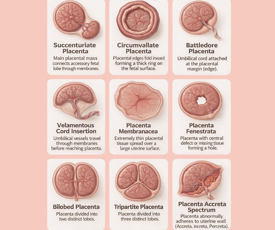
Variations in placental lobes
Sometimes the placenta forms with more than one section, or “lobe”. A bilobed placenta means the placenta has two main sections connected by blood vessels. A tripartite placenta has three lobes instead of one. There is also a variation called a succenturiate lobe, where a small accessory lobe develops separate from the main placenta but remains connected through the membranes. Most of the time these findings do not cause complications, but knowing they are present helps guide monitoring during pregnancy and ensures that the entire placenta is delivered safely after birth.
Differences in cord attachment
Another variation relates to where the umbilical cord attaches. Normally the cord inserts near the centre of the placenta, but sometimes it attaches closer to the edge. This is called a battledore placenta. In a rarer variation known as velamentous cord insertion, the cord vessels travel through the membranes before reaching the placenta rather than being protected within the placental tissue itself. When identified on ultrasound, this can simply mean we monitor the pregnancy a little more closely to ensure blood flow to the baby remains optimal.
Variations in placental shape
Occasionally the placenta develops structural differences in its surface. A circumvallate placenta occurs when the edges of the placenta fold inward, forming a raised ring around the outer edge. Another rare variation is placenta membranacea, where placental tissue spreads thinly across a wider area of the uterus. There are also uncommon findings such as placenta fenestrata, where a small central gap appears within the placental tissue. Many of these are simply interesting anatomical findings rather than something that affects pregnancy significantly.
Placenta accreta spectrum
One placental condition that does require careful planning is placenta accreta spectrum. This occurs when the placenta attaches too deeply into the wall of the uterus. Depending on the depth of attachment, this can be classified as accreta, increta or percreta. Fortunately this is relatively uncommon, but when identified during pregnancy it allows the obstetric team to carefully plan delivery to ensure the safest outcome for both mother and baby.
Why monitoring matters
One of the reasons routine ultrasound scans are so valuable is that they allow us to identify placental position and structure during pregnancy. Most placental variations do not affect the course of pregnancy, but knowing about them helps us tailor care when necessary.
Every pregnancy is unique
The placenta is a temporary but incredibly complex organ, and no two pregnancies are exactly the same. Variations in placental shape, lobes or cord insertion are often simply part of the natural diversity of pregnancy. What matters most is careful monitoring, good communication and a care plan that supports both mother and baby throughout the pregnancy journey.
Dr Abby